"Imagine driving your car in a heavy rainstorm and your wipers don't work. That's what keratoconus does to your eyesight..."-Unknown
Keratoconus has no known cure, and many people do not even know they have it because it begins as nearsightedness and astigmatism. It is a progressive disorder that may progress rapidly or sometimes take years to develop. It can severely affect the way we see the world, including simple tasks such as driving, watching TV, or just reading a book. Some keratoconus patients have described their vision as being "blind with light"
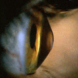
Keratoconus is a non-inflammatory, self-limiting ectasia of the axial portion of the cornea. It is characterized by progressive thinning and steepening of the central cornea. As the cornea steepens and thins, the patient experiences a decrease in vision which can be mild or severe depending on the amount of corneal tissue affected.
|
 |
|
Onset of keratoconus occurs during the teenage years--mean age of onset is age 16 years--but onset has been reported to occur at ages as young as 6 years. Keratoconus rarely develops after age 30 years. Keratoconus shows no gender predilection and is bilateral in over 90% of cases. In general, the disease develops asymmetrically: diagnosis of the disease in the second eye lags about five years after diagnosis in the first. The disease process is active for about five to 10 years, then it may be stable for many years. During the active stage, change may be rapid.
Typically, vision loss can be corrected early by spectacles; later, irregular astigmatism requires optical correction with rigid contact lenses. Contact lenses provide a uniform refracting surface and therefore improve vision. Contact lenses can improve vision, but they can also scar the cornea. Patients should be informed upon diagnosis that they will likely require contact lenses eventually. Although most patients can continue to read and drive, some feel quality of life is adversely affected. Patients need to know that eye examinations will be required annually or more frequently to monitor progression. About 20% of patients will eventually need a corneal transplant.
About Cornea Transplants
In most cases, the surgery is done on an outpatient basis- the patient will enter the hospital or surgery center a few hours prior to surgery and leave that same day- generally a few hours after the surgery. In the pre-op waiting area, the patient will will be prepped, medication will be given to help them relax before surgery. A needle attached to tubing will be inserted to deliver fluids and medications into their vein and EKG leads will be attached to the patient's chest in order to monitor their heart. These are standard safety precautions.
Local or general anesthesia can be used for this procedure. The decision as to which type is used should be discussed with their surgeon preoperatively and is based on their age, general health, length of surgery, their doctor's preference and their anxiety level.
In the operating room, the patient's eyelids are carefully washed and covered with a sterile plastic drape. Oxygen is occasionally provided by a plastic tube placed near the nose. Patients often doze off during the operation, and most are left with vague recollections of a short procedure, although some remember all of it.
The entire procedure is done under a microscope. A circular cookie cutter-like instrument, called a trephine, is used to remove the center of the diseased cornea. A "button" of similar size is cut from the donor cornea. This donor tissue is then sewn in place with extremely fine nylon sutures.
At the conclusion of the procedure, a patch and shield are applied to protect the eye. The patient will then be taken to the recovery room to wait until they are fully awake before being discharged.
Because the cornea has no blood supply, the transplant heals relatively slowly. Sutures are left in place for three months to one year, and in some cases if the vision is good, they are left in permanently. The sutures are buried and therefore don't cause discomfort. Occasionally, they do break and then need to be removed. Often they are removed, adjusted or loosened to improve vision. Suture adjustment and removal are simple, painless office procedures. (Courtesy of NKCF.org)
Information acquired by Center Of Keratoconus Websight (cut and paste is great, isn't it?)
|

